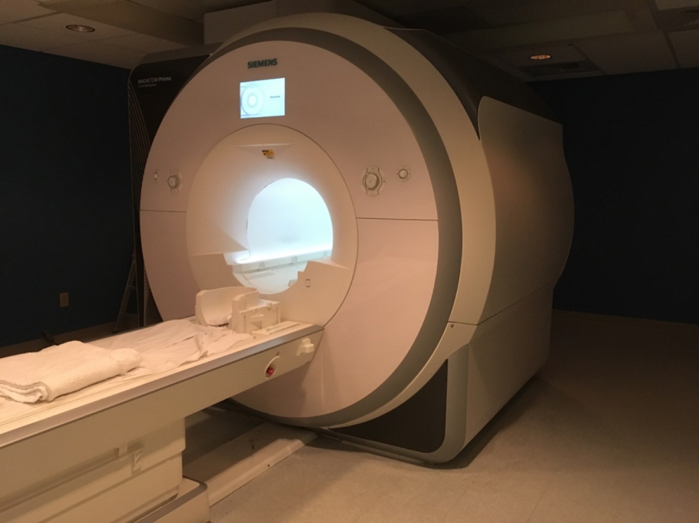
State-of-the-art functional neuroimaging on a Siemens MAGNETOM Prisma 3 Tesla whole-body MRI system, supporting research across the Dartmouth community.
Dartmouth Brain Imaging Center · Directed by Prof. Tor Wager · Open to Dartmouth & external researchersThe DBIC operates a state-of-the-art 3.0 Tesla Siemens MAGNETOM Prisma whole-body MRI system, running on the Siemens XA60 software platform. Its homogeneous 3T magnet, high-performance XR 80/200 gradient system, and Tim 4G integrated-coil architecture deliver excellent B1 homogeneity and enable advanced capabilities such as simultaneous multi-slice acquisition.
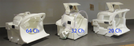
The Tim 4G coil system supports parallel imaging and simultaneous multi-slice acquisition across three receiver coils, chosen to match each study's resolution, coverage, and head-size needs.
| Coil | Best for |
|---|---|
| 64-channel | Highest SNR & iPAT/SMS performance; 40 of 64 channels in the head region |
| 32-channel | iPAT "no-tune" workhorse — parallel imaging & multiband |
| 20-channel | Larger heads; older technology, not generally recommended |
All sequences are customizable — adjust T1/T2, slice thickness, filters, and acceleration to fit your protocol.
T1 MPRAGE, T2, and T2-FLAIR anatomical imaging with high isotropic resolution.
HARDI and multi-shell DWI for tractography and structural connectomics.
Single-band, multiband/SMS, and multi-echo. Standard: 2.7 mm isotropic in <1.5 s/volume.
Non-invasive perfusion imaging for quantitative cerebral blood flow.
Multi-slice MRS (GABA, NAA, and other metabolites), including custom mega-press.
Time-of-flight angiography and field-mapping sequences.
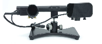
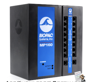
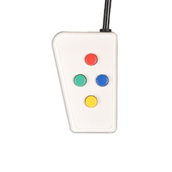
A complete suite for delivering stimuli and recording behavior and physiology, synchronized to scanner acquisition.

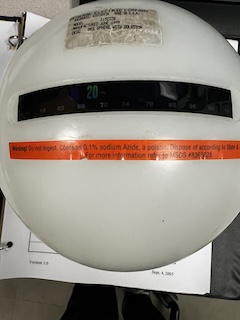
FBIRN QA metrics are generated weekly to monitor scanner stability — mean ghost percentage (threshold <3%), signal-to-fluctuation-noise ratio (SFNR), and SNR — using a standardized fMRI protocol on an agar phantom with the 32- and 64-channel coils. Siemens coil-element checks run weekly for the head coil and monthly for the body coil.
An approved DBIC protocol is required before booking. Scan requests are submitted to Courtney Rogers; all rates include MR tech support, data transfer, and archiving.
| Rate tier | When | Cost |
|---|---|---|
| Internal · Peak | Mon–Fri, 8 a.m.–4 p.m. | $500 / hour |
| Internal · Off-peak | Weeknights after 4 p.m.; weekends | $400 / hour |
| External | Non-Dartmouth-funded projects | $700 / hour |
Among the most cost-effective research rates in the U.S. (typical centers charge $550–$850/hour). Off-peak sessions require a certified operator and scan buddy.
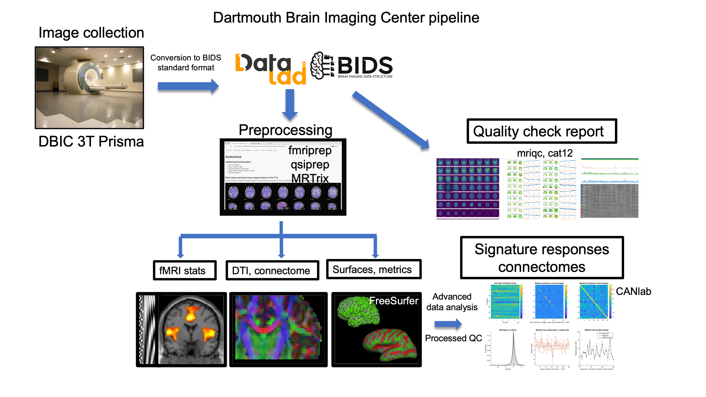
Every study collected at DBIC flows through a standardized, automated pipeline — BIDS conversion and DataLad versioning, preprocessing with fMRIPrep and FreeSurfer, automated quality control with MRIQC, and advanced analysis with CANlab tools. It interoperates with more than 50 BIDS apps.
Explore the pipelineIn 1999, Dartmouth became the first liberal-arts college in the country to own and operate an MRI machine strictly for research. The center opened with a 1.5T GE system, upgraded to a 3.0T Philips in 2004, and has operated its current Siemens 3T Prisma since August 2016. The DBIC is directed by Prof. Tor Wager and staffed by an MRI physicist, an MR technologist, a computer systems manager, and an administrative director.