Three dedicated testing rooms for measuring the body's response to pain, emotion, and social interaction — combining multi-participant autonomic recording, thermal quantitative sensory testing, and video-based behavioral decoding.
Maintained by the Wager Lab (CANlab) · Shared on requestThe Human Psychophysiology & Neuroimaging Laboratory maintains three dedicated testing rooms purpose-built for peripheral physiology and pain research. The rooms are electromagnetically shielded and soundproofed, providing a controlled environment for clean autonomic recordings and for video monitoring that feeds machine-learning models of facial expression and movement.
The facility couples multi-participant BIOPAC recording with a deep suite of somatosensory and pain-testing hardware, supporting everything from single-subject quantitative sensory testing to interpersonal-interaction studies with up to three participants recorded simultaneously.
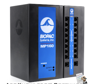
Three BIOPAC MP160 stations record synchronized physiology from up to three participants at once — enabling interpersonal-interaction and hyperscanning-style designs across multiple bodily systems.
Heart rate via electrocardiography (ECG) and photoplethysmography (PPG).
Skin conductance (electrodermal activity) as an index of sympathetic arousal.
Breathing measured with a respiration belt and a nasal cannula.
Trans-radial electrical bioimpedance velocimetry for continuous cardiovascular dynamics.
A FLIR thermal camera captures facial and skin temperature without contact, adding a non-invasive autonomic channel.
Three MP160 stations record multiple people simultaneously for interpersonal-interaction studies.
All channels time-locked for cross-person and cross-system analysis.
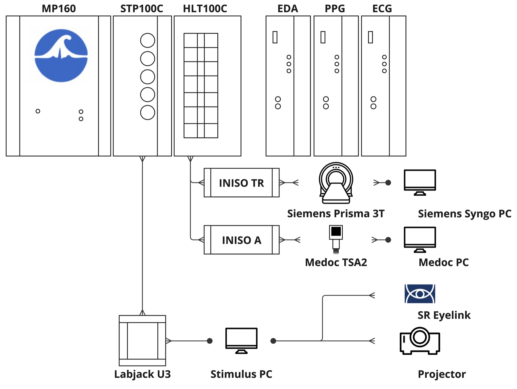
The lab's BIOPAC configuration is designed to interoperate with the wider DBIC ecosystem. Physiology channels can be recorded alongside MRI acquisition on the Siemens Prisma, aligned to thermal stimulation delivered by the Medoc systems, and co-registered with gaze from the EyeLink tracker — so autonomic, neural, and behavioral signals share a common timeline.
The lab houses an unusually deep set of somatosensory stimulators, anchored by six computer-controlled thermal systems for precise heat and cold delivery. Together with custom mechanical devices, they support quantitative sensory testing (QST) and paradigms that probe endogenous pain modulation, including dual stimulation for conditioned pain modulation.
Beyond autonomic physiology, the shielded rooms support video-based behavioral decoding and rapid-setup EEG for combined peripheral-and-central recording.
Continuous video capture in every testing room for objective, camera-based behavioral measurement.
Recordings feed machine-learning models that decode facial expression and body movement.
Electromagnetic shielding and soundproofing keep both physiology and video data clean.
Four 20-channel wet-electrode systems for fast-mounting EEG in dyadic and pain studies.
Rapid-setup electrodes shorten preparation time, easing multi-participant sessions.
Combine EEG, autonomic physiology, pain stimulation, and video within a single paradigm.
The Human Psychophysiology & Neuroimaging Laboratory is part of Tor Wager's Cognitive and Affective Neuroscience Laboratory (CANlab). Lab equipment and facilities are shared on request — researchers interested in dyadic physiology, quantitative sensory testing, or pain paradigms are encouraged to reach out to discuss study design, scheduling, and equipment availability.