A shared facility for non-invasive brain stimulation in research on brain–behavior relationships — pairing MRI-guided transcranial magnetic stimulation, MRI-compatible transcranial electrical stimulation, and steerable temporal-interference stimulation for deep-brain targeting without implants.
Maintained by the Wager Lab · Shared on requestThe Neurostimulation Lab brings together three complementary non-invasive techniques under one roof, so investigators can move from mapping a brain region to directly probing its role in behavior, cognition, and clinical states.
Each system is designed to interoperate with the wider DBIC ecosystem — from MRI-based neuronavigation that targets stimulation to individual anatomy, to an MRI-compatible electrical-stimulation setup verified for use inside the 3T scanner.
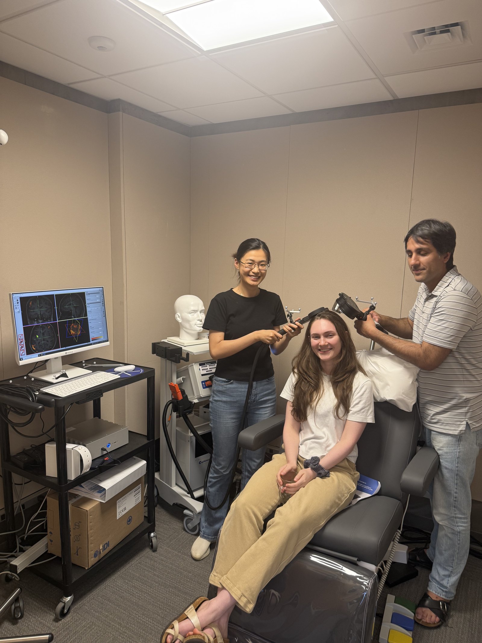
A MagVenture MagPro X100 (230 V) stimulator drives a Cool-B65 A/P fluid-cooled figure-8 coil, which supports both verum (active) and sham stimulation for blinded, controlled designs. Fluid cooling enables the sustained pulse trains needed for repetitive TMS protocols.
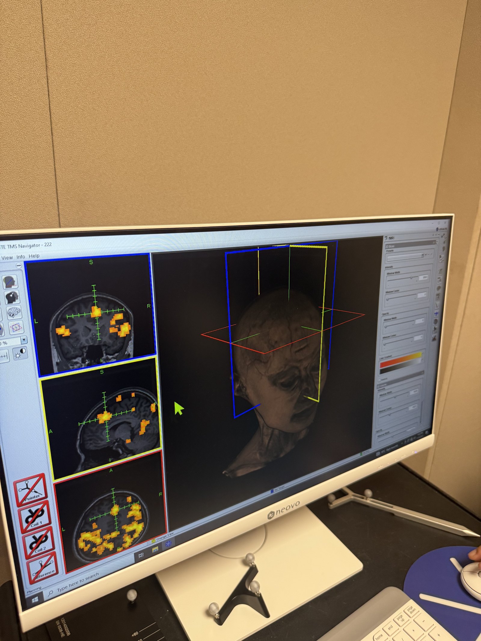
Targeting is guided by the Localite TMS Navigator, an MRI-based neuronavigation system that co-registers each participant's structural scan to their head in real time. Paired with a tracking camera, it lets operators place and maintain the coil over an anatomically or functionally defined target with continuous visual feedback.
| Component | Details |
|---|---|
| Stimulator | MagVenture MagPro X100 (230 V) |
| Coil | Cool-B65 A/P fluid-cooled figure-8, verum & sham |
| Neuronavigation | Localite TMS Navigator, MRI-based |
| Targeting | Real-time optical tracking for continuous coil placement |
Two fMRI-compatible Starstim systems combine transcranial electrical stimulation with concurrent EEG recording at the same electrode sites — letting you stimulate and read out cortical activity together.
Transcranial direct-current stimulation to shift cortical excitability up or down at targeted sites.
Transcranial alternating-current stimulation to entrain and probe rhythmic neural activity.
Transcranial random-noise stimulation for stochastic modulation of cortical responsiveness.
24-bit EEG recorded at the same sites as stimulation, so activity can be monitored throughout a session.
Starstim 8 and Starstim 20 offer a choice of montage density for focal or multi-site protocols.
Designed for use alongside MRI, enabling stimulation studies that pair with functional imaging.
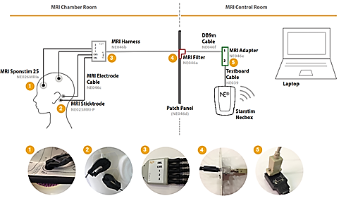
The DBIC operates an MRI-compatible Starstim 8 that can be used with a participant inside the scanner. The stimulator itself sits in the control room, connected to the head coil region through MR-filtered, shielded cables that carry current into the bore without compromising image quality or safety.
Stimulation is delivered through saline-sponge electrodes suited to the MRI environment. The full configuration has been verified as safe for in-scanner use, enabling designs that pair stimulation with functional imaging.
Preparing a tES/tTIS session means fitting a stimulation head cap, placing electrodes at the planned montage sites, and connecting the stimulator control units.
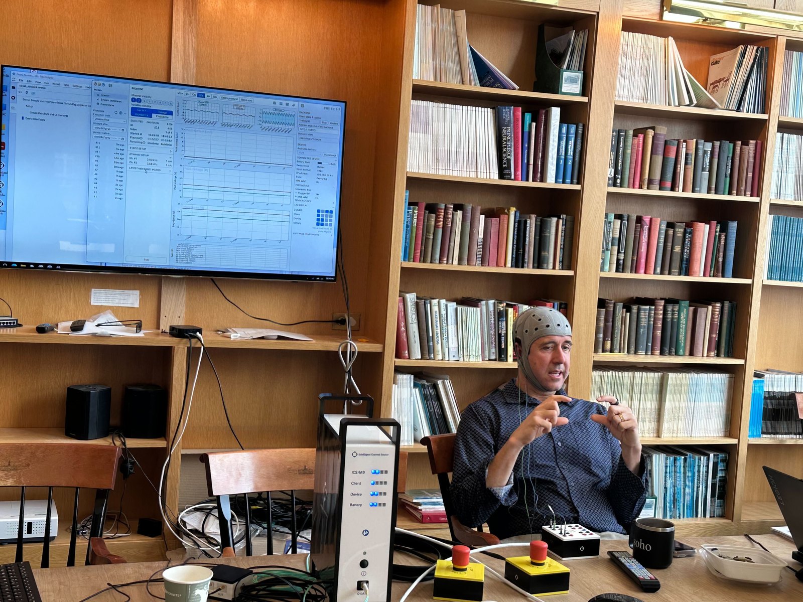
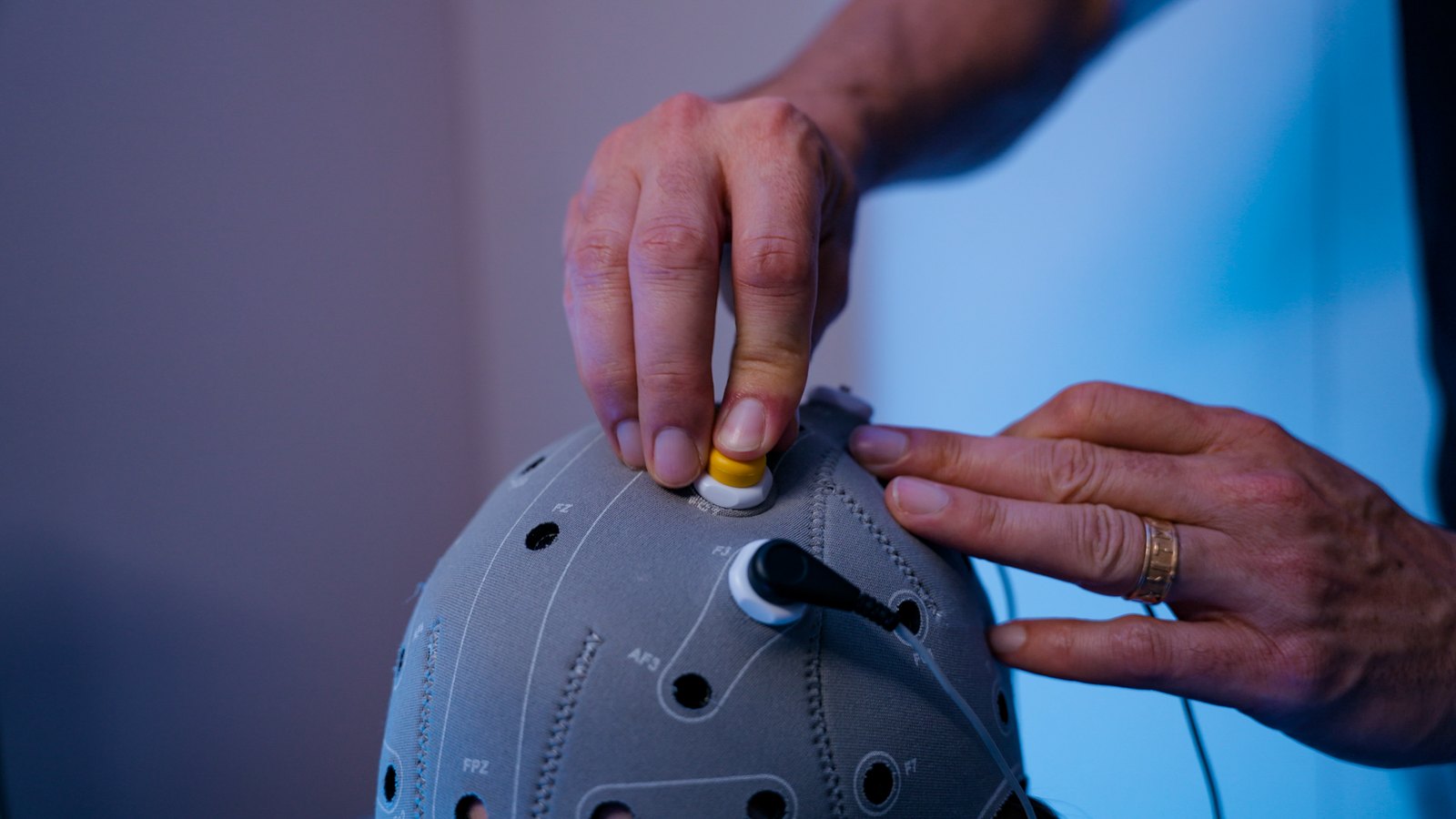
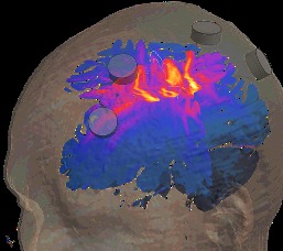
Transcranial temporal interference stimulation (tTIS) is an emerging technique for reaching deep brain structures non-invasively. The lab runs an 8-channel TI Solutions system as an early adopter of the approach.
Rather than stimulating with a single frequency, tTIS applies two current pairs at slightly different kHz-range frequencies. Individually these are too fast to drive neurons, but where the fields overlap they interfere to produce a low-frequency envelope. By adjusting the currents — informed by electric-field modeling of each participant's anatomy — that envelope and the region it modulates can be steered to deep targets without surgical implants.
Every configuration in the lab is operated under verified safety procedures, with particular care in the MRI environment.
The Neurostimulation Lab is maintained by the Wager Lab and shared on request. Because stimulation studies require appropriate training, screening, and protocol approval — and, for in-scanner work, coordination with DBIC scheduling — access is arranged on a per-project basis. Prospective users are encouraged to reach out early to discuss study design, targeting, and safety requirements.