The system
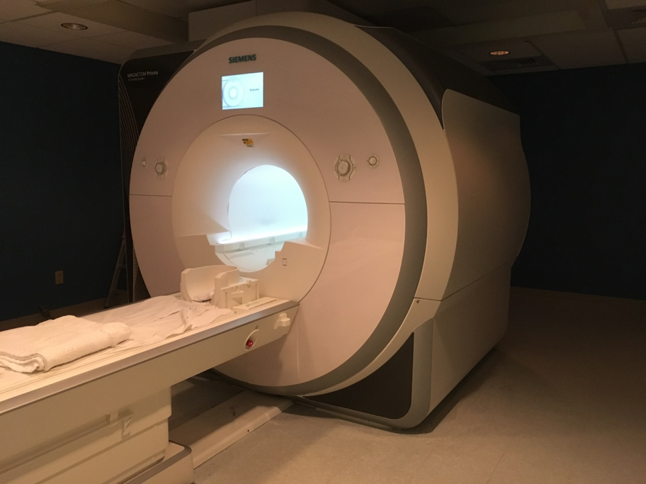
Our center is equipped with a state-of-the-art 3 T Siemens MAGNETOM Prisma whole-body MRI system (Siemens Medical Solutions, Erlangen, Germany) and a variety of peripherals needed for functional neuroimaging.
Software and sequences
The scanner operates on the Siemens XA60 software platform. Vendor supplied packages are available for BOLD (Inline BOLD imaging, 3D PACE syngo and BOLD 3D Evaluation syngo), Diffusion (Diffusion Tensor Imaging, DTI Evaluation and DTI tractography syngo), and Spectroscopy sequences.
Hardware
Advanced hardware integration in our system (e.g. homogeneous 3 T magnet, XR 80/200 gradient system and transmission system) provide excellent B1 homogeneity, and enable novel capabilities such as simultaneous multislice (SMS) acquisition.
Gradients
On our system, the gradient rise time in all three axes is 200 T/m/s and peak gradient strength is 80 mT/m. Such a fast and powerful gradient system provides high-speed structural and functional imaging.
Magnet and shimming
Along with its uniform 3 T magnet, and capability of first and second order shimming, we can acquire high-quality MR spectra and images. The scanner’s magnet delivers excellent magnetic homogeneity of 0.25 ppm, as measured within a 40 cm diameter spherical volume (DSV) in the 60 cm bore.